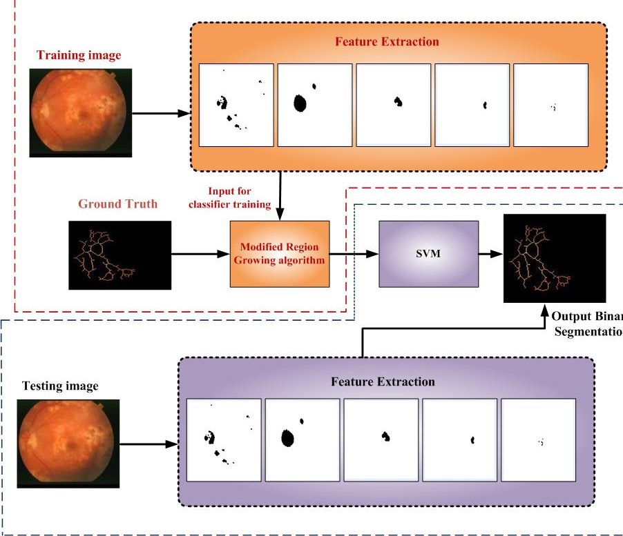


Retinal blood vessels segmentation plays an important role for retinal image analysis. In this code, we propose robust retinal blood vessel segmentation method. A feature is firstly developed to capture local shape information of vessels by employing the length prior of vessels, which is robust to intensity variety. After that, local intensity feature is calculated for each pixel, and then morphological gradient feature is extracted for enhancing the local edge of smaller vessel. At last, line set based feature, local intensity feature, and gradient feature are combined to obtain local descriptions using Modified Region Growing Algorithm. After feature extraction, Support Vector Machine (SVM) is trained for blood vessel segmentation.
Set of Retinal Blood Vessel Images (Testing Images)
Segmentation of the Abnormal Blood Vessel Images (Abnormal Images)
#Retinal, #image, #hypertension, #stroke, #cardiovascular, #capillary pressurizations, #Diabetic, #Retinopathy, #thin, #vessel, #pre-processing, #blood, #vessels, #segmentation, #ophthalmology , #shape, #information, #blurred, #ROI, #eye, #movement, #pupil, #dilation #intensity, #feature, #pixel, #morphological, #gradient, #enhancement, #edge, #Region, #Growing,, #extraction, # SVM, #training, #testing, #abnormal,# human, #eye,
[1]. Karn, P.K., Biswal, B. and Samantaray, S.R., 2018. Robust retinal blood vessel segmentation using hybrid active contour model. IET Image Processing, 13(3), pp.440-450. [2]. Biswal, B., Pooja, T. and Subrahmanyam, N.B., 2017. Robust retinal blood vessel segmentation using line detectors with multiple masks. IET Image Processing, 12(3), pp.389-399.