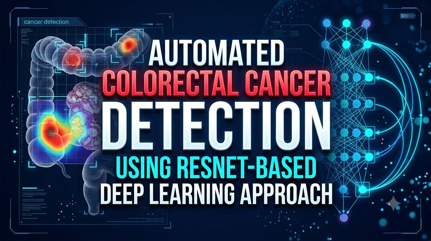
Automated Colorectal Cancer Detection Using ResNet-Based Deep Learning Approach
Colorectal cancer is one of the most common and life-threatening diseases worldwide, contributing significantly to cancer-related mortality. Early detection plays a crucial ...
$ 99.00
Quality Guaranteed
Reviewed project files and clear structure.
24/7 Support
Reach us anytime for setup and usage help.
Instant Access
Download your files right after checkout.
Source Included
Code, assets, and project-ready materials included.
About This Product
Automated Colorectal Cancer Detection Using ResNet-Based Deep Learning Approach
1. Introduction
Colorectal cancer is one of the most common and life-threatening diseases worldwide, contributing significantly to cancer-related mortality. Early detection plays a crucial role in improving patient survival rates and reducing treatment complexity. Traditionally, diagnosis is performed through colonoscopy and histopathological examination, which require expert interpretation and are often time-consuming. These limitations highlight the need for automated and efficient diagnostic systems that can assist medical professionals in accurate decision-making.
Recent advancements in deep learning, particularly in convolutional neural networks (CNNs), have revolutionized the field of medical image analysis. Models such as ResNet50 have demonstrated exceptional performance in extracting complex features from images, making them highly suitable for disease detection tasks. By leveraging transfer learning, pretrained models can be adapted to medical datasets, reducing training time while maintaining high accuracy. This has opened new opportunities for developing intelligent systems for cancer detection.
In this work, a deep learning-based approach is proposed for the classification of colorectal images into three categories: Normal, Polyp, and Serrated adenoma. The methodology includes data preprocessing techniques such as resizing, normalization, and contrast enhancement using CLAHE, followed by data augmentation to balance the dataset. A ResNet50-based architecture is then employed for feature extraction and classification, ensuring robust and reliable performance across different image conditions.
Furthermore, the trained model is integrated into a web-based application using Django, enabling real-time image upload and prediction. This end-to-end system provides a user-friendly interface for automated diagnosis, making it practical for real-world applications. The proposed approach not only improves diagnostic efficiency but also reduces human error, thereby contributing to early detection and better management of colorectal cancer.
2. Problem Statement
Manual detection of colorectal abnormalities is:
- Time-consuming
- Prone to human error
- Dependent on expert availability
There is a need for an automated, reliable, and efficient system that can classify colorectal images into categories such as Normal, Polyp, and Serrated adenoma with high accuracy.
3. Input
·Medical image dataset (colorectal images)
·Image formats: .jpg, .png, .jpeg
·Classes:
- Normal
- Polyp
- Serrated adenoma
·User input (in web app):
- Uploaded colorectal image
4. Output
·Predicted class label:
- Normal
- Polyp
- Serrated adenoma
·Confidence score (percentage)
·Visual display of uploaded image with prediction result
5. Novelty
·Use of ResNet50 pretrained model for improved feature extraction
·Integration of CLAHE (Contrast Limited Adaptive Histogram Equalization) for enhanced image quality
·Balanced dataset using data augmentation
·End-to-end system combining:
- Deep Learning model
- Django web interface
·Real-time prediction through a web application
6. Contribution
·Developed a complete pipeline:
- Data preprocessing
- Augmentation
- Model training
- Evaluation
- Deployment
·Implemented:
- Multi-class classification system
- Performance evaluation metrics:
- Accuracy
- Precision
- Recall
- F1-score
- ROC-AUC
·Built a user-friendly web interface using Django:
- Login/Signup system
- Image upload
- Instant prediction
7. Conclusion
The proposed system successfully automates colorectal cancer detection using a ResNet50-based deep learning model. The model achieves high accuracy and provides reliable classification results. Integration with a Django-based web application makes the system practical for real-world usage. This approach reduces dependency on manual diagnosis and supports early detection, which can significantly improve patient outcomes.
8. Reference
[1] J. Urban, P. Tripathi, T. Alkayali, M. Mittal, F. Jalali, W. Karnes, and P. Baldi, “Deep Learning-Based Classification of Colorectal Polyps on Whole-Slide Images,” The Lancet Gastroenterology & Hepatology, vol. 3, no. 6, pp. 1–10, 2018.
[2] J. N. Kather, A. Halama, and A. Marx, “100,000 Histological Images of Human Colorectal Cancer and Healthy Tissue,” PLOS Medicine, vol. 16, no. 5, 2019.
[3] G. Litjens, T. Kooi, B. E. Bejnordi, et al., “A Survey on Deep Learning in Medical Image Analysis,” Medical Image Analysis, vol. 42, pp. 60–88, 2017.
[4] K. He, X. Zhang, S. Ren, and J. Sun, “Deep Residual Learning for Image Recognition,” in Proceedings of the IEEE Conference on Computer Vision and Pattern Recognition (CVPR), 2016.
[5] K. Simonyan and A. Zisserman,“Very Deep Convolutional Networks for Large-Scale Image Recognition,” in International Conference on Learning Representations (ICLR), 2015.
Backend Programming Language
Python
Backend Web Framework
Django
Frontend Structure
HTML
Frontend Styling
CSS
No reviews available for this product.